
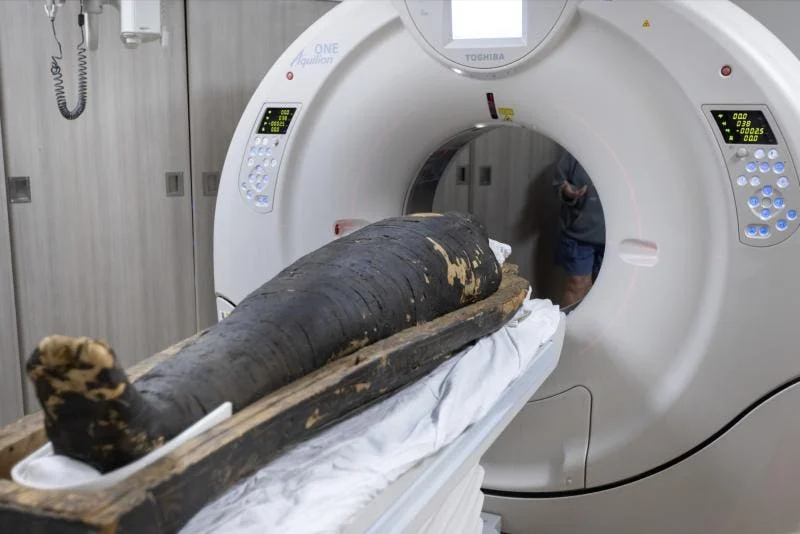
In the sterile, fluorescent-lit corridors of Keck Medicine at the University of Southern California, a patient unlike any other recently made his way through the aperture of a high-end CT scanner. He did not arrive with a contemporary medical record or a list of modern symptoms. Instead, he arrived swaddled in blackened linen shrouds, his biography written in the desiccated tissues and mineralized bones of a life that ended more than 2,300 years ago. This patient, an ancient Egyptian priest named Nes-Hor, represents the vanguard of a new era in archaeology—one where the scalpel is replaced by the X-ray beam, and the "unwrapping" of history occurs in a purely digital realm.
The recent study of Nes-Hor and a fellow priest, Nes-Min, marks a significant milestone in the field of paleopathology. By utilizing state-of-the-art 320-slice computed tomography (CT) technology, researchers are now able to peer through the millennia with unprecedented clarity, uncovering medical secrets that were previously obscured by the limitations of 20th-century imaging. These findings do more than just satisfy historical curiosity; they provide a vital link in our understanding of the evolution of human disease, the history of surgical intervention, and the enduring biological legacy of the ancient world.
The Evolution of the Digital Autopsy
For centuries, the study of mummified remains was a destructive process. Early "mummy unwrapping" events in the 19th century were often public spectacles that resulted in the permanent loss of anatomical and archaeological context. It was not until the advent of radiography that scientists could glimpse the interior of a mummy without disturbing its physical integrity. However, early X-rays provided only flat, two-dimensional shadows, often resulting in a jumble of overlapping bones and resin that was difficult to interpret.

The introduction of CT scanning in the late 20th century revolutionized the field, allowing for cross-sectional "slices" of the body. Nes-Min and Nes-Hor were actually scanned once before, in the 1990s. Yet, the resolution of that era was akin to looking through a fogged window. Today’s technology has cleared that glass. The 320-slice scanner used at USC offers a resolution of approximately 0.5 millimeters—a level of detail that allows radiologists to identify tiny fractures, dental abscesses, and even potential tool marks from ancient medical procedures.
This transition from physical to digital archaeology is not merely a change in toolsets; it is a shift in philosophy. The "digital twin" of a mummy allows researchers across the globe to collaborate on a single specimen without ever touching the original remains. It preserves the sanctity of the deceased while maximizing the scientific yield, turning the mummy into a permanent, searchable database of ancient biological information.
Clinical Profiles from the Ptolemaic Era
The results of the recent scans have provided remarkably specific diagnoses for the two priests. Nes-Min, who lived around 330 BCE and died in his forties, was revealed to have suffered from significant spinal degeneration. The scans showed a collapsed lumbar vertebra and broken ribs, conditions that likely caused him chronic, debilitating pain. In the context of the Ptolemaic era, a man in his forties was entering the later stages of life, and his skeletal remains tell a story of a body taxed by both time and perhaps the physical requirements of his station.
Most intriguing, however, were the anomalies found in Nes-Min’s skeletal structure. Summer Decker, the head of 3D imaging for Keck Medicine of USC, noted the presence of holes and marks that do not appear to be natural or the result of post-mortem damage. These "tool marks" suggest the possibility of early surgical intervention. If confirmed, this would be a monumental discovery, providing physical evidence of sophisticated neurosurgical or orthopedic attempts in ancient Egypt—a civilization already known for its advanced, albeit rudimentary, medical papyri.

Nes-Hor, who died nearly a century and a half later, around 190 BCE, lived into his sixties—a venerable age for the time. His scans revealed the heavy toll of longevity. He suffered from severe dental issues and a hip joint so deteriorated that he would have required a staff or the assistance of others to move. These findings paint a poignant picture of the lived experience in antiquity. We often view ancient figures through the lens of mythology or monumental architecture, but the CT scanner reminds us that they were biological entities who suffered from the same wear and tear, the same osteoarthritis, and the same toothaches that plague modern humans.
The Technology: 320 Slices of History
The technical prowess required to image a mummy is vastly different from that required for a living patient. Living tissue is hydrated and possesses varying densities that are easy for X-rays to distinguish. Mummies, by contrast, are extremely dry and often filled with resins, linen, and amulets that can create "artifacts" or distortions in the digital image.
The 320-slice scanner overcomes these hurdles by taking hundreds of images as the gantry—the doughnut-shaped housing of the X-ray tube—rotates around the subject. By capturing so many data points, the computer can reconstruct a three-dimensional volume that is accurate to a fraction of a millimeter. This allows for "segmentation," a process where researchers can digitally remove the linen wrappings, the skin, or the resin to isolate a single bone or organ.
At USC, this data was used to create life-sized 3D-printed models of the priests’ anatomy. Seeing a digital model is one thing; holding a 1:1 reproduction of a 2,300-year-old fractured spine allows for a tactile understanding of the pathology. This bridge between the digital and the physical is becoming a standard in high-end archaeological research, providing a way for museums to share their findings with the public in a way that is both educational and deeply respectful.

Paleopathology and the Modern Health Landscape
The study of mummies like Nes-Hor and Nes-Min contributes to a broader field known as paleopathology—the study of ancient diseases. In 2024, a massive study involving CT scans of over 200 mummies from various eras revealed a surprising prevalence of atherosclerotic heart disease and stroke. This challenged the long-held belief that "lifestyle diseases" are a purely modern phenomenon driven by processed foods and sedentary habits.
By examining the remains of individuals who lived thousands of years ago, scientists can track the genetic and environmental history of diseases like cancer, diabetes, and heart disease. If a priest in 330 BCE had the same arterial clogging as a modern office worker, it suggests that some predispositions to disease are baked into human biology, regardless of the era. The detailed scans of Nes-Min’s potential surgical sites also offer a timeline for the history of medicine, showing how early humans attempted to mitigate the pain and suffering revealed by their bones.
Ethics, Preservation, and the Future of Archaeology
As technology advances, the ethical considerations of scanning human remains become more complex. There is a growing movement toward "virtual repatriation," where the digital data of a mummy is returned to its country of origin, allowing local scholars to lead the research. The use of non-invasive CT scans is widely considered the most ethical way to study these individuals, as it avoids the desecration of the body while still allowing for the advancement of knowledge.
Looking forward, the next frontier in this field is the integration of Artificial Intelligence (AI). AI algorithms are currently being trained to recognize specific patterns in ancient bone density and tissue desiccation that might be missed by the human eye. We may soon see "automated paleopathology," where a computer can scan a mummy and instantly provide a full medical history, comparing it against a global database of thousands of other specimens.

Furthermore, advancements in "Spectral CT" imaging—which uses different energy levels of X-rays to identify the chemical composition of materials—could allow researchers to identify the specific types of resins, oils, and herbs used in the mummification process. This would provide a chemical map of ancient trade routes and embalming techniques that are currently invisible to standard scanners.
Conclusion: A Window into the Human Condition
The exhibition of Nes-Min and Nes-Hor at the California Science Center’s "Mummies of the World" exhibit is a testament to the power of modern technology to bridge the gap between the present and the deep past. As Diane Perlov, an anthropologist at the center, noted, these technologies offer a "powerful window" into lives that would otherwise be lost to the sands of time.
When we look at the 3D-rendered face of Nes-Hor, we are not looking at a relic; we are looking at a person. We see his struggle with a failing hip, his discomfort from dental decay, and the care that was taken to preserve his body for eternity. The CT scanner has done more than unlock medical secrets; it has restored the humanity of these ancient individuals. In the 0.5-millimeter slices of a 320-slice scan, we find the universal story of the human body—its fragility, its resilience, and its enduring quest for healing. As technology continues to evolve, the silent priests of Egypt will continue to speak, offering us a clearer vision of where we came from and, perhaps, a better understanding of where our biological future is headed.